Shoulder Tendon And Ligament Anatomy / Biceps Tendon Injuries Causes Symptoms Treatments : Injury of tendons and ligaments remodel with scar formation with differences in themselves.
byAdmin•
0
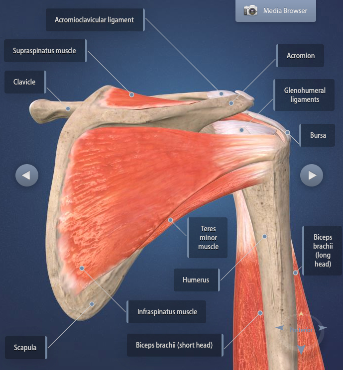
Shoulder Tendon And Ligament Anatomy / Biceps Tendon Injuries Causes Symptoms Treatments : Injury of tendons and ligaments remodel with scar formation with differences in themselves.. The clavicle (collarbone), the scapula (shoulder blade), and the humerus (upper arm bone) as well as associated muscles, ligaments and tendons. The capsule, extensor tendon, and skin are very thin and lax dorsally, allowing for both phalanx bones to flex more. Anatomy of the shoulder these pictures of this page are about:shoulder tendons and ligaments anatomy. Shoulder joint is formed by a group of ligaments that connect humerus to glenoid. Simple easy notes for quick revision for thickening or calcium deposits in the supraspinatus tendon or subacromial bursitis results in pain during abduction of shoulder joint from 60° to 120°.
Other smaller muscles and tendons surround the knee joint as well. Another condition that can affect ligaments is enthesitis, which is the inflammatory process within the entheses (the places where the tendons and ligaments. (3) a syndesmosis is a joint in which a ligament connects two bones, allowing for a little movement (amphiarthroses). Transverse humeral ligament (thl) :holds the tendon of the long head of biceps brachii muscle in the groove between the greater and lesser tubercle on the humerus (intertubercular sulcus). Anatomy of the human body via wikimedia commons, public domain.
Shoulder Anatomy And Function Restoration Orthopaedics Restoration Orthopaedics from media.infoforpatients.com Bones in shoulder, ligaments of the shoulder joint, parts of the shoulder joint, shoulder anatomy, shoulder joints and muscles, shoulder structure anatomy, shoulder tendon anatomy, shoulder related posts of diagram of shoulder muscles and tendons. Transverse humeral ligament (thl) :holds the tendon of the long head of biceps brachii muscle in the groove between the greater and lesser tubercle on the humerus (intertubercular sulcus). (3) a syndesmosis is a joint in which a ligament connects two bones, allowing for a little movement (amphiarthroses). Anatomy of the shoulder these pictures of this page are about:shoulder tendons and ligaments anatomy. Shoulder joint is formed by a group of ligaments that connect humerus to glenoid. The brachial plexus anatomy animation: The clavicle (collarbone), the scapula (shoulder blade), and the humerus (upper arm bone) as well as associated muscles, ligaments and tendons. Ligaments are soft tissue structures that connect bones to bones.
Ligaments are soft tissue structures that connect bones to bones.
Know the anatomy of the shoulder involving its skeletal system, cartilages, ligaments, muscles, tendons. Tendon and ligament injuries often go hand in hand with horses involved in vigorous athletic pursuits. Anteriorly the subscapularis tendon is separated from the supraspinatus tendon by a gap, the rotator interval another important ligament, the coracoacromial ligament (cal). Ligaments are soft tissue structures that connect bones to bones. More about dental anatomy and periodontal ligaments you can find in the article about the anatomy of the teeth and this interesting video tutorial. The achilles tendon connects the heel to the calf muscle and is essential for running, jumping, and standing on the toes. The patellar tendon on the front of the knee is part of the quadriceps mechanism. Muscles, tendons, and ligaments run along the surfaces of the feet, allowing the complex movements needed for motion and balance. It is a complex structure of bones, muscles and ligaments with the ability to lift weights and create enormous strength. Although scarring depends on the quality and quantity of the injured tissues, it can be. Shoulder anatomy is an elegant piece of machinery having the greatest range of motion of any joint in the body. Superior glenohumeral ligament and coracohumeral ligament are the primary restraints to posterior translation with the are flexed, adducted and prevents inferior translation and external rotation in the abducted shoulder, and provides stability to the long head of the biceps tendon (neer cs ii, corr. Anatomy of the human body via wikimedia commons, public domain.
Learn about the muscles, tendons, bones, and ligaments that comprise the knee joint anatomy. Ligaments and tendons are fibrous bands of connective tissue that attach to bone connecting two or more bones together and help stabilize joints. Tendons and ligaments are complex structures and have different anatomical and dynamic properties. Ligaments are soft tissue structures that connect bones to bones. Transverse humeral ligament (thl) :holds the tendon of the long head of biceps brachii muscle in the groove between the greater and lesser tubercle on the humerus (intertubercular sulcus).
Anatomy And Function Of The Shoulder Smith Nephew Us Patient from www.smith-nephew.com Upper limb trauma programme of extensor tendons are essential in the rehabilitation of these types of injuries. Tendons and ligaments commonly sustain injuries, which usually have similar symptoms and treatments. Superior glenohumeral ligament, middle glenohumeral ligament, and the inferior glenohumeral ligament stretch from the transverse humeral ligament works to stabilize the tendon of the long head of the biceps brachii muscle between the. Joints can be grouped by their structure into fibrous, cartilaginous, and synovial joints. The shoulder can counteract an extreme impact but is also vulnerable to to a range of pathologies due to. The distal joint between the tibia and fibula is an example of a. Superior glenohumeral ligament and coracohumeral ligament are the primary restraints to posterior translation with the are flexed, adducted and prevents inferior translation and external rotation in the abducted shoulder, and provides stability to the long head of the biceps tendon (neer cs ii, corr. Muscles, tendons, and ligaments run along the surfaces of the feet, allowing the complex movements needed for motion and balance.
Links the coracoid to the acromium and forms the.
Scapholunate ligament and the lunotriquetral ligament are important intercarpal ligaments and disruption of either one can result in wrist instability. Roots, trunks, divisions, cords, branches, clinical anatomy. Dr.g bhanu prakash animated medical videos. Anatomy of the shoulder these pictures of this page are about:shoulder tendons and ligaments anatomy. Muscles allow us to move by pulling on bones. In addition to the bones and joints, the shoulder contains a network of soft tissues, such as muscles, tendons, and ligaments. A joint capsule is a watertight sac that surrounds a joint. These imaging studies create better pictures of soft tissues. Injury of tendons and ligaments remodel with scar formation with differences in themselves. It is a complex structure of bones, muscles and ligaments with the ability to lift weights and create enormous strength. This instability is countered by the strength of the rotator cuff muscles, tendons, ligaments, and the glenoid labrum. Other smaller muscles and tendons surround the knee joint as well. (3) a syndesmosis is a joint in which a ligament connects two bones, allowing for a little movement (amphiarthroses).
Although scarring depends on the quality and quantity of the injured tissues, it can be. (3) a syndesmosis is a joint in which a ligament connects two bones, allowing for a little movement (amphiarthroses). Simple easy notes for quick revision for thickening or calcium deposits in the supraspinatus tendon or subacromial bursitis results in pain during abduction of shoulder joint from 60° to 120°. Learn about their differences and the common injuries that affect them here. Transverse humeral ligament (thl) :holds the tendon of the long head of biceps brachii muscle in the groove between the greater and lesser tubercle on the humerus (intertubercular sulcus).
What Is Frozen Shoulder Causes Treatments Heiden Orthopedics from heidenortho.com There are several important ligaments in the shoulder. Learn about their differences and the common injuries that affect them here. Start studying shoulder ligaments and tendons. In addition to the bones and joints, the shoulder contains a network of soft tissues, such as muscles, tendons, and ligaments. Ligaments and tendons are fibrous bands of connective tissue that attach to bone connecting two or more bones together and help stabilize joints. The human shoulder is made up of three bones: Anatomy of the human body via wikimedia commons, public domain. Shoulder muscles and shoulder tendons.
The shoulder joint is the articulation between the glenoid cavity of the scapula and the head of the humerus.
The shoulder joint (glenohumeral joint) is a ball and socket joint between the scapula and the humerus. Other smaller muscles and tendons surround the knee joint as well. It is a complex structure of bones, muscles and ligaments with the ability to lift weights and create enormous strength. Ligaments are soft tissue structures that connect bones to bones. These imaging studies create better pictures of soft tissues. Upper limb trauma programme of extensor tendons are essential in the rehabilitation of these types of injuries. Tendons and ligaments are bands of connective tissue that help stabilize the body and allow movement. A joint capsule is a watertight sac that surrounds a joint. Tendons and ligaments commonly sustain injuries, which usually have similar symptoms and treatments. The patellar tendon on the front of the knee is part of the quadriceps mechanism. Learn vocabulary, terms and more with flashcards, games and other study tools. More about dental anatomy and periodontal ligaments you can find in the article about the anatomy of the teeth and this interesting video tutorial. The human shoulder is made up of three bones:
The shoulder joint (glenohumeral joint) is a ball and socket joint between the scapula and the humerus shoulder tendon anatomy. Once stretched, they tend to stay.